Protecting Against Inferior Calcaneal Spur

Overview
Heel spurs are a small calcification, which extends into the attachment of the connective tissues from the calcaneus (heel bone). This is easily observed on X-Ray and is very commonly misdiagnosed as the cause of your pain. Why? Because the wrong type of scan is used and only one foot is examined. Do you know if you have a spur on the other heel, the heel without the pain? Probably? and the heel spur would have been there long before you had the pain, and will still be there long after the pain is gone. Heel spurs are not the actual cause of heel pain, and are virtually always a secondary observation or symptom caused by long term pulling on the heel bone.
Causes
Fctors that increase the risk of developing heel spurs include a high body mass index (BMI), regular vigorous activity, and intensive training routines or sports. Factors such as these are believed to increase the incidence of repetitive stress injuries that are associated with the formation of heel spurs. When a heel spur forms, extremely sharp pain along with the feeling that a part of the heel is trying to burst through the skin usually occurs. If left untreated, an individual may eventually begin to struggle to perform simple activities such as walking.
Symptoms
It is important to be aware that heel spurs may or may not cause symptoms. Symptoms are usually related to the plantar fasciitis. You may experience significant pain and it may be worse in the morning when you first wake up or during certain physical activities such as, walking, jogging, or running.
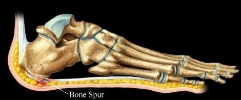
Diagnosis
Diagnosis of a heel spur can be done with an x-ray, which will be able to reveal the bony spur. Normally, it occurs where the plantar fascia connects to the heel bone. When the plantar fascia ligament is pulled excessively it begins to pull away from the heel bone. When this excessive pulling occurs, it causes the body to respond by depositing calcium in the injured area, resulting in the formation of the bone spur. The Plantar fascia ligament is a fibrous band of connective tissue running between the heel bone and the ball of the foot. This structure maintains the arch of the foot and distributes weight along the foot as we walk. However, due to the stress that this ligament must endure, it can easily become damaged which commonly occurs along with heel spurs.
Non Surgical Treatment
Treatments for bone spurs and plantar fasciitis include Stretching the calf muscles several times daily is critical in providing tension relief for the plantar fascia. Some physicians may recommend using a step to stretch, while others may encourage yoga or pushing against a wall to stretch. Icing after activity. A frozen tennis ball can provide specific relief. Rolling the tennis ball under the arch of the foot after exercise can lessen pain in the area. Taping is also recommended at times. Several manufacturers of sports tape have plantar fascia specific lines. Orthotics are a good idea for those on their feet during the day. Orthotics can provide cushioning and relief. Cortisone shots in the fascia can provide temporary anti-inflammatory relief. Losing weight is perhaps the most effective method of improving heel and foot pain. Those who are overweight are far more likely to report these syndromes.
Surgical Treatment
Surgery to correct for heel spur syndrome is a common procedure which releases plantar fascia partially from its attachment to the calcaneous (heel bone). This part of the surgery is called a plantar fasciotomy due to the fact the fascia is cut. This is most often done through an open procedure as any heel spur or bursa can be removed at the same time. If the spur is not removed during the surgery, it will probably be just as successful, as the large spur is not the true problem. Some physicians use an endoscopic approach (EPF) where a small camera aids the physician during surgery with typically smaller incisions on each side of your foot.